Ocular Trauma

Eye Injuries

Eye Trauma

Eye trauma refers to damage caused by a direct blow to the eye. The trauma may affect not only the eye, but the surrounding area, including adjacent tissue and bone structure.

There are many different forms of trauma, varying in severity from minor injury to medical emergencies. Even in cases where trauma seems minor, every eye injury should be given medical attention.

What Causes Eye Trauma?

When the eye is hit with blunt force, it suddenly compresses and retracts. This can cause blood to collect underneath the hit area, which leads to many of the common symptoms of eye trauma.

Symptoms of Eye Trauma

Symptoms of eye trauma may include:

  • Pain
  • Trouble seeing
  • Cuts to the eyelid
  • One eye not moving as well 
  • One eye sticks out
  • Blood in the clear part of the eye
  • Unusual pupil size or shape
  • Something embedded in the eye
  • Something under the eyelid that cannot be easily removed

Treatments for Eye Trauma

Every eye injury should be given medical attention; do not touch, rub or try to remove any object in the eye. If the eye has been cut or there is an object in the eye, rest a protective shield – such as a paper cup – on the bone around your eye. Make sure there is no pressure on the eye itself. Seek immediate, professional medical attention.

In minor cases of trauma, such as a black eye from a sports injury, applying cold to the affected area can help bring swelling down, and allow the affected area to heal faster. However, even in cases where trauma seems minor, every eye injury should be given medical attention.

The best way to avoid eye trauma is to prevent it by using protective eyewear while doing things that may put them at risk. Activities include home repair, yard work, cleaning, cooking, and playing sports. In most cases of injury, people report not properly protecting their eyes – which shows that proper precautions may prevent an eye injury.

Light and your eyes

Explore the latest research regarding bright lights and retina damage, and how to properly protect your eyes.

Parents will often tell children not to stare at the sun. This is good advice, since permanent retinal damage occurs after staring for just a few minutes! This is called solar retinopathy. Lasers and very bright industrial lights can also damage the retina.

Since very bright lights can damage the retina in a short period of time, can long-term exposure to moderately bright light cause retinal damage? The answer is maybe, and why not protect yourself against this possibility?

Research Findings on Bright Lights and Eye Damage

In the Chesapeake Bay Waterman Study, which analyzed fisherman exposed to bright light reflected off the water every day, blue light exposure was found to increase the risk of age-related macular degeneration (AMD). This conclusion, while suggestive, as not definitive, since it is difficult to quantify light exposure in everyday life. 

In experimental mice, bright light does cause permanent retinal damage. If the light has the intensity of sunlight, short exposure times can cause damage. If the light is not quite so bright, chronic exposure over days to weeks can cause permanent damage. This is thought to be due to what is called photo-oxidative damage; the light reacts with the retina to produce molecules that are very reactive and cause damage to surrounding molecules.

If light exposure is a risk factor for AMD, then it would be expected that people with lighter colored eyes, which let in more light, would have a higher risk. Indeed, African-Americans have a much lower risk of AMD than Caucasians. Some studies have suggested that people with blue eyes have the highest risk.

Some experimental mice and even a particular breed of dog have genetic mutations that make their retinas extremely sensitive to light; permanent retinal damage can be caused by light levels that would not harm animals with normal retinas. Analogously, people with AMD, Stargardt disease, or retinitis pigmentosa may be more susceptible to retinal light damage than people with normal retinas.

How Can I Protect My Eyes from Bright Lights?

The simplest way to protect against possible retinal light damage is to wear sunglasses and a hat. The sunglasses should have dark tint. If you hold the glasses up to a light, the light coming through the lenses should appear grey, brown, or yellowish brown—not blue, which is probably the most damaging light wavelength. If picking ready-made sunglasses in a store, choose the ones that transmit the least light (but only wear them if you can still see well enough).  If ordering custom sunglasses, ask the optician for a very dark tint. Polarization will also decrease the amount of light coming through sunglasses and helps reduce glare. UV protection helps protect the lens inside the eye against cataracts. Since UV light is blocked by the lens inside the eye it is the visible light that can harm the retina. 

Since it is thought that blue light can damage the retina, ophthalmologists can now offer blue-blocking lens implants when performing cataract surgery. While these may be helpful, the same effect can be achieved by wearing sunglasses.

Are Indoor Lights Damaging to the Eyes?

Could indoor lights be harmful? It is prudent to use the amount of light you need to read or perform activities of daily life, but there is no reason to exceed this amount with very bright lighting. When choosing light bulbs, those with the “warm” color spectrum, favoring reds and greens over the “cool” blues, are typically more comfortable for reading and may be safer. Energy-efficient LED bulbs come in warm or full spectrum varieties. As with most things in life, light is good, in moderation

Joshua Dunaief, MD, PhDScheie Eye Institute, University of Pennsylvania

Transposition of ophthalmic lenses

Transposition

by Andrew Othuke Akpeli

Introduction / definition

According to Efe (2012) transposition as the process of changing a lens prescription from one form to another equivalent form without  changing the value i.e. it is also  to rewrite the expression of its power without actually changing its value. It can also be define as the act of converting the prescription of an ophthalmic lens form a sphere with minus cylinder form to a sphere with plus cylinder form or vice vise. Example – 3D sphere – 2D cylinder axis 180 transpose to – 5D sphere + 2D cylinder axis 90 prescription consists of the sphere, cylinder and the axis. The sphere correct hyperopic and myopia, the cylinder corrects the amount of astigmatism and the axis shows where the stigmatism correction is located.

Types of transposition

Flat Transposition

Toric transposition

Flat transposition: This is the process of transposition whereby the cylinder power and spherical power is separated by 90. The flat transposition is use in the dispensing laboratory.

Toric transposition: This is the process of transposition whereby the cylinder power and spherical power is not separated by 90. The toric transposition is use in the production laboratory.

Lens prescription

Lens prescription can be written in any of this form

  1. Plano cylinder
  2. Sphero cylinder
  3. Cross cylinder

Plano cylinder: This has one surface plane and the other, either + or – cylinder eg (plano x 0.50DCx180)

Sphere cylinder:  This has one surface spherical and the other cylindrical eg +0.75DS -1.00DC x 50

Cross cylinder: This has one surface a cylinder and other another cylinder eg +1.50DCx180/0.75DCx90

Rules of transposition

  1. Transposition of sphero-cylindrical prescription from plus to minus cylinder (or vice versa)

Rules

Step 1: New spherical power: add the power of the sphere to the power of cylinder (algebraically) of a given prescription

Step 2: New cylindrical power: change the sign of the cylinder (plus or minus to the opposite sign) with same power.

Step 3: New axis: add or subtract 90 from the given axis (depending on which value is higher) i.e. if is below 90, add 90 to it but if is above 90 subtract 90. Let us now apply this rule to a specific sphero – cylindrical prescription.

  1. Given + 1.00DS – 1.00DC x 90

Related Topic  The Ministry of Infrastructure (MININFRA) ,22 JOBS Vacancies in Rwanda

From the on set, it is important to note that this prescription has been written in minus cylinder from. To convert it to plus cylinder form, we apply the rules as follows.

Rule 1: (new spherical power); (+1.00) + (1.00) = plano

Rule 2: (new cylindrical power) change the cylinder sign with same value = +1.00DC

Rule 3: (new axis) add or subtract 90 (depending on which value is greater) i.e if is below 90, add 90 to it but if it is above 90 subtract 90. 90 + 90 = 180. Therefore, the transposed plus cylindrical form of a given prescription is plano + 1.00DC x 180

  1. Given -2.00DS + 1.50DC x 45

To transpose this prescription from plus to its minus cylindrical form we apply same rules as follows:

Rule 1: (new sphere power) – 2.00 + 1.50 = – 0.50DS

Rule 2: (new cylinder power) change the cylinder sight with the same power -1.50DC

Rules 3: (New axis) i.e if is below 90, add 90 to it but if is above 90 subtract 90 45 + 90 = 135

Thus, the transpose form of this prescription into its minus cylindrical form is equivalent to :- 0.50DS – 135

Transposition of sphero-cylinders into cross cylinders

A sphero-Cylindrical prescription can be broken down into two cross-cylinder that it was originally made of this form of. This form of transposition is also guided by its set of rules

Rules

Rule 1. (1st Cross-Cylinder): Take the spherical power of a given sphero-cylinder prescription and write this as a cross cylinder with its axis 90 away from the axis of a given sphero-cylindrical prescription.

Rule 2. (2nd cross-cylinder) add the spherical and cylindrical power of a given sphero-cylindrical prescription and write this as cross cylinder with its axis same as the axis of the sphero-cylindrical prescription.

Rules 3. (both cross-cylinder) combine the two cross cylinder obtain from rule 1 and rule 2 above as the equivalent cross cylinder for the sphero-cylindrical prescription.

 The follow are some examples to illustrate the application of the rules

  • Given -4.00DS – 0.50DC x 90

Related Topic  Connected Talent,IT Resourcer/Candidate Manager Jobs in Australia

Rule 1. (1st cross-cylinder) -4.00DC x 180

Rule 2. (2nd cross-cylinder) -4.50DC x 90

Rule 3. (3rd cross-cylinder) +4.00DC x 180/-4.50DC x 90

  • Given: -1.50DS + 0.75DC x 45

Rule 1. (1st cross-cylinder) -1.00DC x 135

Rule 2. (2nd cross-cylinder) -0.75DC x 45

Rule 3. (3rd cross-cylinder) +1.50DC x 135/-075DC x 45

Transposition of 2 cross-cylinder into sphero cylinder

Rule 1: (sphere power) take either power of the 2 cross-cylinders as spherical power of the new sphero-cylinders as spherical power of the new sphero-cylinderical prescription.

Rule 2: (cylindrical power) subtract the power of the cylindrical chosen as sphere from the power of the second cross-cylinder and make this the cylinder of the new sphero-cylinder

Rule 3: (axis) take the axis of the cross-cylinder that was not chosen as sphere power as axis of the new sphero-cylinderical prescription

The following are some examples to illustrate application of the rule

  1. Given: +2.00DS x 180/+1.00DC x 90

Rule 1. (sphere power) + 2.00DS

Rule 2. (cylindrical power) +1.00 – (+2.00) = -1.00DC

Rule 3. (axis): 90

Therefore, the sphero-cylindrical prescription is: +2.00DC – 1.00DC x 90

  1. Given: -1.00DC x 135/-3.00DCx45

Rule 1. (sphere power) -3.00DS

Rule 2. (cylindrical power) -1.00 – (-3.00)

-1.00 + 3.00 = +2.00

Rule 3. (axis): 135

Therefore. The sphero-cylindrical prescription is: -3.00DC + 2.00DC x 135

Toric transposition

A toric lens is practical same as a cylindrical lens. By these design the surfaces of toric lenses are more curved (or bent) than the surfaces of cylindrical lenses. But unlike the flat cylindrical lens prescription, the toxic prescription will indicate the various curves that have been incorporated during the process of production to arrive at a particular prescription. This explain why a toric lens prescription have the following three parts

  1. The base curve (BC)
  2. The cross curve (CC)
  3. The sphere curve (SC)

To produce a particular prescription from a lens blanks these three components with their different powers will be generated in the factory. This process is called surfacing

Since most of the physical properties of toric lenses are similar to cylindrical lens and vice versa. This means that given a specific toric lens prescription, the sphero-cylindrical prescription of equivalent power can be deduced from it by applying the following rules.Related Topic  procreteconstruction 3 people Job Vacancy in Australia

Rule 1 (sphere power) = base curve + sphere curve

Rule 2 (cylinder power) = cross curve – base curve

Rule 3 (axis) same as axis of cross curve

The following example is meant to illustrate the application of those rules:

Rule 1 (sphere power): +2.00 + (-3.00) = -1.00DS

Rule 2 (cylinder power) +4.00 – 2.00 = +2.00DC

Rules 3 (axis) = 180

Sphero-cylindrical prescription: -1.00DS + 2.00DC x 180

Importance of transposition

After an optometrist has done a refraction for a patient, the optometrist takes the prescription to the optician and the lens prescription is given to be + 1.00DS-2.00DC x 180 and the optician check the stocks of lens available and founds that (+1.00DS – 2.00DC x 180) prescription is not in stock, what the optician need to do is to transpose the prescription.

+1.00DS – 2.00DC x 180

– 1.00DS + 2.00DC x 90.

So instead of the optician finding it as a problem of getting + 1.00DS-2.00DC x 180 the optician simply go for -1.00DS + 2.00DC x90 it makes dispensing cylindrical lenses easy instead of keeping your patient  waiting or ordering for more lenses when unknowing to you it is your stock.

Conclusion

In conclusion, transposition is important because it helps and optician to get an equivalent (same) prescription for a given prescription. It is also important for an optician to be acquitted with the knowledge of transposition. An optician should not be ignorant of keeping a patient waiting or placing an order for a cylindrical lens prescription when there is an alternative form of that prescription in stock, all you need to do is to transpose.

References

Efe Odjimogho, O.D. FNCO (2012). In laboratory manual, senior lecturer optometry department faulty of life science, university of Benin.

Mr. Nsubuisi Igboekwe (2013). Ophthalmic workshop dispensing lab ii

San Francisco, American Academy of ophthalmology 2011-2012

Allied Health on the job training kit 2011

Dallas American Health Association Academy of ophthalmology 2011 (Revised as appropriate)FacebookTwitterEmailWhatsAppPinterest

What Is Type 2 Diabetes?

Type 2 Diabetes

What Is Type 2 Diabetes?

Type 2 diabetes is a lifelong disease that keeps your body from using insulin the way it should. People with type 2 diabetes are said to have insulin resistance.

People who are middle-aged or older are most likely to get this kind of diabetes. It used to be called adult-onset diabetes. But type 2 diabetes also affects kids and teens, mainly because of childhood obesity.

Type 2 is the most common type of diabetes. There are about 29 million people in the U.S. with type 2. Another 84 million have prediabetes, meaning their blood sugar (or blood glucose) is high but not high enough to be diabetes yet.

Signs and Symptoms of Type 2 Diabetes

The symptoms of type 2 diabetes can be so mild that you don’t notice them. About 8 million people who have it don’t know it. Symptoms include:

  • Being very thirsty
  • Peeing a lot
  • Blurry vision
  • Being cranky
  • Tingling or numbness in your hands or feet
  • Fatigue/feeling worn out
  • Wounds that don’t heal
  • Yeast infections that keep coming back
  • Feeling hungry
  • Weight loss without trying
  • Getting more infections

If you have dark rashes around your neck or armpits, see your doctor. These are called acanthosis nigricans, and they can be signs that your body is becoming resistant to insulin.CONTINUE READING BELOW

Causes of Type 2 Diabetes

Your pancreas makes a hormone called insulin. It helps your cells turn glucose, a type of sugar, from the food you eat into energy. People with type 2 diabetes make insulin, but their cells don’t use it as well as they should.

At first, your pancreas makes more insulin to try to get glucose into your cells. But eventually, it can’t keep up, and the glucose builds up in your blood instead.

Usually, a combination of things causes type 2 diabetes. They might include:

  • Genes. Scientists have found different bits of DNA that affect how your body makes insulin.
  • Extra weight. Being overweight or obese can cause insulin resistance, especially if you carry your extra pounds around your middle.
  • Metabolic syndrome. People with insulin resistance often have a group of conditions including high blood sugar, extra fat around the waist, high blood pressure, and high cholesterol and triglycerides.
  • Too much glucose from your liver. When your blood sugar is low, your liver makes and sends out glucose. After you eat, your blood sugar goes up, and your liver will usually slow down and store its glucose for later. But some people’s livers don’t. They keep cranking out sugar.
  • Bad communication between cells. Sometimes, cells send the wrong signals or don’t pick up messages correctly. When these problems affect how your cells make and use insulin or glucose, a chain reaction can lead to diabetes.
  • Broken beta cells. If the cells that make insulin send out the wrong amount of insulin at the wrong time, your blood sugar gets thrown off. High blood sugar can damage these cells, too.

CONTINUE READING BELOW

Type 2 Diabetes Risk Factors

Certain things make it more likely that you’ll get type 2 diabetes. The more of these that apply to you, the higher your chances of getting it are. Some things are related to who you are:

  • Age. 45 or older
  • Family. A parent, sister, or brother with diabetes
  • Ethnicity. African American, Alaska Native, Native American, Asian American, Hispanic or Latino, or Pacific Islander American

Risk factors related to your health and medical history include:

Other things that raise your risk of diabetes have to do with your daily habits and lifestyle. These are the ones you can do something about:

CONTINUE READING BELOW

Type 2 Diabetes Diagnosis and Tests

Your doctor can test your blood for signs of type 2 diabetes. Usually, they’ll test you on 2 days to confirm the diagnosis. But if your blood glucose is very high or you have many symptoms, one test may be all you need.

  • A1c. It’s like an average of your blood glucose over the past 2 or 3 months.
  • Fasting plasma glucose. This is also known as a fasting blood sugar test. It measures your blood sugar on an empty stomach. You won’t be able to eat or drink anything except water for 8 hours before the test.
  • Oral glucose tolerance test (OGTT). This checks your blood glucose before and 2 hours after you drink something sweet to see how your body handles the sugar.

Type 2 Diabetes Treatment

Managing type 2 diabetes includes a mix of lifestyle changes and medication.

Lifestyle changes

You may be able to reach your target blood sugar levels with diet and exercise alone.

  • Weight loss. Dropping extra pounds can help. While losing 5% of your body weight is good, losing at least 7% and keeping it off seems to be ideal. That means someone who weighs 180 pounds can change their blood sugar levels by losing around 13 pounds. Weight loss can seem overwhelming, but portion control and eating healthy foods are a good place to start.
  • Healthy eating. There’s no specific diet for type 2 diabetes. A registered dietitian can teach you about carbs and help you make a meal plan you can stick with. Focus on:
  • Eating fewer calories
  • Cutting back on refined carbs, especially sweets
  • Adding veggies and fruits to your diet
  • Getting more fiber
  • Exercise. Try to get 30 to 60 minutes of physical activity every day. You can walk, bike, swim, or do anything else that gets your heart rate up. Pair that with strength training, like yoga or weightlifting. If you take a medication that lowers your blood sugar, you might need a snack before a workout.
  • Watch your blood sugar levels. Depending on your treatment, especially if you’re on insulin, your doctor will tell you if you need to test your blood sugar levels and how often to do it.

CONTINUE READING BELOW

Medication

If lifestyle changes don’t get you to your target blood sugar levels, you may need medication. Some of the most common for type 2 diabetes include:

CONTINUE READING BELOW

Even if you change your lifestyle and take your medicine as directed, your blood sugar may still get worse over time. That doesn’t mean you’ve done something wrong. Diabetes is progressive, and many people eventually need more than one drug.

When you take more than one drug to control your type 2 diabetes, that’s called combination therapy.

You and your doctor should work together to find the best mix for you. Usually, you’ll keep taking metformin and add something else.

What that is may depend on your situation. Some drugs control blood sugar spikes (your doctor may call this hyperglycemia) that come right after meals, for instance. Others are more effective at stopping drops in blood sugar (hypoglycemia) between meals. Some may help with weight loss or cholesterol, as well as your diabetes.

You and your doctor should talk about any possible side effects. Cost may be an issue as well.

If you take medication for something else, that will need to be factored into any decision.CONTINUE READING BELOW

You’ll need to see your doctor more often when you start taking a new combination of drugs.

You might find that adding a second drug doesn’t bring your blood sugar under control. Or the combination of two drugs might work only for a short time. If that happens, your doctor might consider a third noninsulin drug, or you may start insulin therapy.

Type 2 Diabetes Prevention

Adopting a healthy lifestyle can help you lower your risk of diabetes.

  • Lose weight. Dropping just 7% to 10% of your weight can cut your risk of type 2 diabetes in half.
  • Get active. Thirty minutes of brisk walking a day will cut your risk by almost a third.
  • Eat right. Avoid highly processed carbs, sugary drinks, and trans and saturated fats. Limit red and processed meats.
  • Quit smoking. Work with your doctor to keep from gaining weight after you quit, so you don’t create one problem by solving another.

CONTINUE READING BELOW

Type 2 Diabetes Complications

Over time, high blood sugar can damage and cause problems with your:

  • Heart and blood vessels. You’re up to five times more likely to get heart disease or have a stroke. You’re also at high risk of blocked blood vessels (atherosclerosis) and chest pain (angina).
  • Kidneys. If your kidneys are damaged or you have kidney failure, you could need dialysis or a kidney replacement.
  • Eyes. High blood sugar can damage the tiny blood vessels in the backs of your eyes (retinopathy). If this isn’t treated, it can cause blindness.
  • Nerves. This can lead to trouble with digestion, the feeling in your feet, and your sexual response.
  • Skin. Your blood doesn’t circulate as well, so wounds heal slower and can become infected.
  • Pregnancy. Women with diabetes are more likely to have a miscarriage, a stillbirth, or a baby with a birth defect.
  • Sleep. You might develop sleep apnea, a condition in which your breathing stops and starts while you sleep.
  • Hearing. You’re more likely to have hearing problems, but it’s not clear why.
  • Brain. High blood sugar can damage your brain and might put you at higher risk of Alzheimer’s disease.
  • Depression. People with the disease are twice as likely to get depressed as people who don’t have it.

The best way to avoid these complications is to manage your type 2 diabetes well.

  • Take your diabetes medications or insulin on time.
  • Check your blood sugar.
  • Eat right, and don’t skip meals.
  • See your doctor regularly to check for early signs of trouble.

CONTINUE READING BELOW

Build Your Health Care Team

There are many medical professionals who can help you live well with diabetes, including:

  • Endocrinologists
  • Nurses
  • Registered dietitians
  • Pharmacists
  • Diabetes educators
  • Foot doctors
  • Eye doctors
  • Dentists

10 Questions to Ask Your Doctor About Diabetes

If you were recently diagnosed with type 2 diabetes, ask your doctor these questions at your next visit.

  1. Does having diabetes mean that I am at higher risk for other medical problems?
  2. Should I start seeing other doctors regularly, such as an eye doctor?
  3. How often should I test my blood sugar, and what should I do if it is too high or too low?
  4. Are there any new medications that I could use to help manage my diabetes?
  5. Does diabetes mean I have to stop eating the foods I like best?
  6. How can exercise make a difference in my diabetes?
  7. If I’m overweight, how many pounds do I have to lose to make a difference in my health?
  8. Are my children at increased risk for the disease?
  9. What is the importance of diet in diabetes?
  10. Do I need to take my medications even on days that I feel fine?

Glare Reducing Lenses: Understanding Their Uses

Glare Reducing Lenses: Understanding Their Uses

There are two main types of glare reducing lenses for eyewear: lenses with an anti-reflective coating and polarized lenses. Both help to prevent glare in their own way. Glare reducing lenses can improve vision clarity, help people see better while driving at night, reduce annoying glare from water or other horizontal surfaces, and eliminate noticeable reflections on a lens itself.

What is glare?

Glare is caused by light bouncing off of a reflective surface. When talking about eyewear, people are most likely referring to lens glare or environmental glare. Lens glare is caused by the reflection of light off the surface of a lens. Whether it belongs to a camera, telescope, binoculars, or even just glasses, all lenses have some level of reflection with the lowest amount of reflection being less than 0.1%. Eyeglass lenses without a glare-reducing coating typically allow around 90% of light to pass through, depending on the lens material. The other 10% of the light reflects off the surfaces of the lens. The glare caused by this 10% reduces vision clarity, causes people to see halos around headlights and street lamps at night, and creates bright, almost white reflections on the lens itself.

Environmental glare is caused by light waves reflecting off of flat surfaces like water or the highway. It becomes focused and travels in a uniform direction parallel to that surface, creating a bright and intense reflection that we call glare. This type of glare affects everyone, regardless of whether or not they wear glasses.

glare reducing lenses

Outline, in Black

How do glare reducing lenses work?

While it may be impossible to eliminate 100% of the glare on glasses lens, technology has helped to get the number as close to 0 as possible. While both anti-reflective coatings and polarized lenses help to reduce glare, the technology behind these two is quite different. An anti-reflective coating (also known as AR or anti-glare coating) actually encourages more light to pass through a lens. When more light passes through, less light reflected off its surfaces, and thus, less glare.

Polarized lenses, on the other hand, reduce glare by absorbing light waves from a certain orientation. Most polarized lenses for eyewear are oriented to absorb horizontal light waves reflected off of flat surfaces like a lake or the snow-covered ground.

When it comes to eyewear, AR coatings are applied both eyeglass lenses and sunglass lenses. Anti-glare coating is applied to both sides of a lens to prevent light from reflecting off the back of the lens as well. Polarized lenses are typically used for sunglasses since the nature of its glare reducing technology is to block light instead of letting more through.

The benefits of glare reducing lenses

Many people question whether or not it’s worth it to get glare reducing lenses. The short answer is: while not everyone may need sunglasses with polarized lenses, lenses with an anti-reflective coating will vastly improve the quality of life for a glasses wearer.

Lens glare is a major source of eye strain since it reduces vision clarity, forcing your eyes to work harder to focus. People who work with computers are especially susceptible to this type of eye strain since illuminated screens act as a direct and constant source of glare on lenses. Adding AR coating to your lenses significantly lessens this glare, helps you see more clearly, and reduces eye strain caused by computer screens.

Glares called “halos” can be seen around the headlights of cars and street lamps. These halosare a great source of discomfort and distraction for glasses wearers who drive at night. They reduce visibility and make nighttime driving difficult. Anti-glare coating prevents these halos and helps to make driving at night safer for glasses wearers.

If you’re someone who is both literally and figuratively in the spotlight a lot, anti-reflective coating is a must. Glare caused by bright lights reflecting off a lens can be distracting. It also obscures your eyes, making it harder for people to find direct eye contact with you. So if you have a client- or customer-facing job, make sure to consider getting glare reducing glasses.

Finally, if you’re someone who spends a lot of time out on the water or working in the snow, you’re well aware of how much glare can reduce visibility and make it a literal pain to be outside. The tint on sunglasses with polarized lenses helps to reduce that all around brightness, while the polarization helps to save your eyes from blinding glare.

So for the best comfort while wearing glasses or sunglasses, consider glare reducing lenses. Not only will they help you see better, but they’ll also help you get the most out of life. At EyeBuyDirect, you can find affordable glare reducing lenses for any of our great styles.

Coined from eyebuydirect.com

Don’t let glaucoma steal your vision

Overview

Open-angle glaucoma
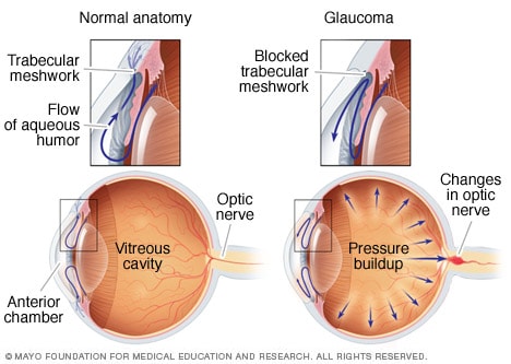
Open-angle glaucomaOpen pop-up dialog box

Glaucoma is a group of eye conditions that damage the optic nerve, the health of which is vital for good vision. This damage is often caused by an abnormally high pressure in your eye.

Glaucoma is one of the leading causes of blindness for people over the age of 60. It can occur at any age but is more common in older adults.

Many forms of glaucoma have no warning signs. The effect is so gradual that you may not notice a change in vision until the condition is at an advanced stage.

Because vision loss due to glaucoma can’t be recovered, it’s important to have regular eye exams that include measurements of your eye pressure so a diagnosis can be made in its early stages and treated appropriately. If glaucoma is recognized early, vision loss can be slowed or prevented. If you have the condition, you’ll generally need treatment for the rest of your life.

Symptoms

The signs and symptoms of glaucoma vary depending on the type and stage of your condition. For example:

Open-angle glaucoma

  • Patchy blind spots in your side (peripheral) or central vision, frequently in both eyes
  • Tunnel vision in the advanced stages

Acute angle-closure glaucoma

  • Severe headache
  • Eye pain
  • Nausea and vomiting
  • Blurred vision
  • Halos around lights
  • Eye redness

If left untreated, glaucoma will eventually cause blindness. Even with treatment, about 15 percent of people with glaucoma become blind in at least one eye within 20 years.

REQUEST AN EYE EXAMINATION AT EYEUPDATE CLINIC

When to see a doctor

Promptly go to an emergency room or an eye doctor’s (ophthalmologist’s) office if you experience some of the symptoms of acute angle-closure glaucoma, such as severe headache, eye pain and blurred vision.

Causes

Glaucoma is the result of damage to the optic nerve. As this nerve gradually deteriorates, blind spots develop in your visual field. For reasons that doctors don’t fully understand, this nerve damage is usually related to increased pressure in the eye.

Elevated eye pressure is due to a buildup of a fluid (aqueous humor) that flows throughout the inside of your eye. This internal fluid normally drains out through a tissue called the trabecular meshwork at the angle where the iris and cornea meet. When fluid is overproduced or the drainage system doesn’t work properly, the fluid can’t flow out at its normal rate and eye pressure increases.

Glaucoma tends to run in families. In some people, scientists have identified genes related to high eye pressure and optic nerve damage.

Types of glaucoma include:

Open-angle glaucoma

Open-angle glaucoma is the most common form of the disease. The drainage angle formed by the cornea and iris remains open, but the trabecular meshwork is partially blocked. This causes pressure in the eye to gradually increase. This pressure damages the optic nerve. It happens so slowly that you may lose vision before you’re even aware of a problem.

Angle-closure glaucoma

Angle-closure glaucoma, also called closed-angle glaucoma, occurs when the iris bulges forward to narrow or block the drainage angle formed by the cornea and iris. As a result, fluid can’t circulate through the eye and pressure increases. Some people have narrow drainage angles, putting them at increased risk of angle-closure glaucoma.

Angle-closure glaucoma may occur suddenly (acute angle-closure glaucoma) or gradually (chronic angle-closure glaucoma). Acute angle-closure glaucoma is a medical emergency.

Normal-tension glaucoma

In normal-tension glaucoma, your optic nerve becomes damaged even though your eye pressure is within the normal range. No one knows the exact reason for this. You may have a sensitive optic nerve, or you may have less blood being supplied to your optic nerve. This limited blood flow could be caused by atherosclerosis — the buildup of fatty deposits (plaque) in the arteries — or other conditions that impair circulation.

Glaucoma in children

It’s possible for infants and children to have glaucoma. It may be present from birth or develop in the first few years of life. The optic nerve damage may be caused by drainage blockages or an underlying medical condition.

Pigmentary glaucoma

In pigmentary glaucoma, pigment granules from your iris build up in the drainage channels, slowing or blocking fluid exiting your eye. Activities such as jogging sometimes stir up the pigment granules, depositing them on the trabecular meshwork and causing intermittent pressure elevations.

REQUEST AN EYE EXAMINATION AT EYEUPDATE CLINIC

Risk factors

Because chronic forms of glaucoma can destroy vision before any signs or symptoms are apparent, be aware of these risk factors:

  • Having high internal eye pressure (intraocular pressure)
  • Being over age 60
  • Being black, Asian or Hispanic
  • Having a family history of glaucoma
  • Having certain medical conditions, such as diabetes, heart disease, high blood pressure and sickle cell anemia
  • Having corneas that are thin in the center
  • Being extremely nearsighted or farsighted
  • Having had an eye injury or certain types of eye surgery
  • Taking corticosteroid medications, especially eyedrops, for a long time

Prevention

These self-care steps can help you detect glaucoma in its early stages, which is important in preventing vision loss or slowing its progress.

  • Get regular dilated eye examinations. Regular comprehensive eye exams can help detect glaucoma in its early stages, before significant damage occurs. As a general rule, the American Academy of Ophthalmology recommends having a comprehensive eye exam every five to 10 years if you’re under 40 years old; every two to four years if you’re 40 to 54 years old; every one to three years if you’re 55 to 64 years old; and every one to two years if you’re older than 65. If you’re at risk of glaucoma, you’ll need more frequent screening. Ask your doctor to recommend the right screening schedule for you.
  • Know your family’s eye health history. Glaucoma tends to run in families. If you’re at increased risk, you may need more frequent screening.
  • Exercise safely. Regular, moderate exercise may help prevent glaucoma by reducing eye pressure. Talk with your doctor about an appropriate exercise program.
  • Take prescribed eyedrops regularly. Glaucoma eyedrops can significantly reduce the risk that high eye pressure will progress to glaucoma. To be effective, eyedrops prescribed by your doctor need to be used regularly even if you have no symptoms.
  • Wear eye protection. Serious eye injuries can lead to glaucoma. Wear eye protection when using power tools or playing high-speed racket sports in enclosed courts.

Health benefits of bitter kola


Did you know that Coca-Cola got its name from a common African tree?

Some of the first recipes for Coca-Cola were made using the extract of the bitter kola plant. Though the company hasn’t used actual kola to flavor their sodas in years, the name remains a reminder of the unusual plant that inspired the iconic drink.

Bitter kola, also known as bitter cola or Garcinia Kola, is a plant found in Central and Western Africa that has long been valued for its medicinal properties. Although traditional African medicine uses all parts of the Bitter Kola plant, the seeds are mostly commonly eaten.

Bitter kola seeds have a sharp, bitter flavor that eases into a slight sweetness as you chew, and they’re typically eaten raw.

Health Benefits

Although bitter kola has been eaten in Africa for years, scientists are just beginning to study the health benefits of this flowering plant.

According to these early studies, bitter kola may be able to help with health problems including:CONTINUE READING BELOW

Infections

Bitter kola has been used over the years to fight infections from the common cold to hepatitis. A 2018 study showed that bitter kola can help combat coughs, bacterial infections, and viral infections. Eating bitter kola when an infection starts may help fight the infection and make you feel better more quickly.

Inflammation

Bitter kola seeds have commonly been chewed on in Africa to fight against inflammatory conditions like arthritis. One 2008 study showed that patients with osteoarthritis in their knees showed significantly reduced inflammation when eating bitter kola compared to a placebo. The high levels of potassium in bitter kola may be a contributing factor in reducing inflammation.

Diabetes

Early studies suggest that a chemical called kolaviron, which is found in bitter kola, may protect against hypoglycemia in people with type 2 diabetes. Although this first study was done on rats and has not yet been reproduced on humans, these early findings are promising.

Nutrients per Serving

The FDA considers bitter kola generally safe to eat, but classifies it similarly to essential oils. That means there’s not much information available about serving sizes or nutritional data. However, a 2013 study found that bitter kola was high in the following nutrients:

  • Carbohydrates
  • Fat
  • Protein

The study same study also found that bitter kola had high levels of:

  • Vitamin C
  • Calcium 
  • Potassium
  • Iron
  • Caffeine

How to Prepare Bitter Kola

Bitter kola can be eaten on its own, uncooked, like many other nuts. Keep in mind, however, that it’s called “bitter” for a reason. Bitter kola can be an acquired taste.

You can sometimes find drinks made or flavored with bitter kola at African markets and online stores. If you don’t like the taste but want to introduce bitter kola to your diet, these drinks may be a good option.

Road safety and your eyesight

Uncorrected vision continues to endanger the lives of drivers, passengers, and pedestrians around the world. Compounded by external factors such as nighttime driving, inclement weather, and adverse road conditions, uncorrected vision contributes to more than 1.25 million road accident deaths each year.[1] Until recently, the intersection of vision and road safety has not received adequate attention. Consequently, there is a lack of awareness around the need to address the impact of vision problems on driver and road safety. While governments and policymakers can play a significant role in raising  greater awareness of this public health threat, eye care professionals are also part of the solution as they are a competent authority in promoting effective assessment of vision and correcting drivers’ visual functions.CONTENTKEY TAKEAWAYSREFERENCES

The problematic relationship between uncorrected vision and road safety is a deadly, global phenomenon, and is growing more dangerous. The World Health Organization (WHO) has reported that road traffic crashes are a leading cause of road deaths globally and the main cause of deaths among those aged 15 to 29 years-old.[2] What is perhaps most disturbing is how traffic accidents disproportionately affect low- or middle-income countries, which are home to more than 80 percent of the world’s population. Though these countries account for only 54 percent of the world’s vehicles, they see 90 percent of all road traffic deaths. These road accidents also come with an adverse economic impact, estimated at US$500 billion a year –another burden on low- or middle-income countries. [2]

“These road accidents also come with and adverse economic impact, estimated at US$ 500 Billion a year – another burden on low- or middle-income countries.”

There are hopeful signs, yet much work remains. Globally, the number of traffic deaths plateaued between 2007 and 2015 despite a four percent increase in the world’s population – and an increase in motorization even four times higher than that – suggesting that intervention efforts can and do save lives. [2] However, disparity remains. Among the 68 countries that saw a rise in the number of road traffic deaths between 2010 and 2013, 84 percent were low- or middle-income.[2]

Recognizing the severity and disparity of both the human toll and economic burden that traffic accidents place on national economies and individual households alike, the United Nations, in its Sustainability Goals, included an ambitious goal of halving traffic-related deaths and injuries by the year 2020.

This new imperative to address road safety brings with it a greater urgency to understand the underlying causes of traffic accidents. Much attention has been given to the effects of drunk driving and, more recently, the use of wireless devices while driving. But in addition to the important issues of inebriated and inattentive driving, vision impairment also warrants attention.

The assumption of good vision, necessary to safely operate a vehicle, has long caused uncorrected vision to be overlooked as a factor in road safety. Drs. Cynthia Owsley and Gerald McGwin note in their analysis, “Vision and Driving,” that “the visual demands of driving are intricate.” [3] By cataloging the many and varied visual tasks involved in driving, including the simultaneous use of central and peripheral vision to monitor primary and secondary tasks, they reinforce how visually intensive driving under normal conditions is. These findings have led researchers to conclude that many visual tests for drivers are inadequate, often failing to simulate the distractions and wide ranging contrast and luminance levels experienced in real-world road conditions.[3] This is compounded by the fact that many drivers avoid seeking evaluation and treatment for vision issues; in Europe, 19 percent of drivers reported delaying visits to an optician until they notice problems with their vision.[5]

Driving and vision infographics Vision Impact Institute - Points De Vue

The contributions of Owsley, McGwin, Chakrabarty and others to the growing dialogue around driving and vision are indicative of the increased attention and analysis that this issue requires. However, though the link between vision and road safety should be a global concern, the disparity between high-income and low- or medium-income countries illustrates that it remains very much a local issue, which demands a greater understanding of local factors.

Road Safety Around the World

As the correlation between vision and road safety has come to the forefront, more data is becoming available from both high-income and low- or medium-income countries.

In India, for example, the dual forces of population growth and economic development have resulted in a higher number of vehicles on the road and, consequently, a greater number of traffic accidents. One study of vision and drivers in India calculated the road crash involvement rate of drivers with unacceptable vision test results at 81 percent, which was 30 percent higher than that of drivers with good vision.[6] While many factors, including poor vehicle and road conditions and traffic violations, have been found to affect driver safety, researchers have pointed to vision problems,  worsened by night driving, as significant causes of driver safety challenges.

As the correlation between vision and road safety has come to the forefront, more data is becoming available from both high-income and low- or medium-income countries.

In India, for example, the dual forces of population growth and economic development have resulted in a higher number of vehicles on the road and, consequently, a greater number of traffic accidents. One study of vision and drivers in India calculated the road crash involvement rate of drivers with unacceptable vision test results at 81 percent, which was 30 percent higher than that of drivers with good vision.6 While many factors, including poor vehicle and road conditions and traffic violations, have been found to affect driver safety, researchers have pointed to vision problems,  worsened by night driving, as significant causes of driver safety challenges.

Driving and vision infographics Vision Impact Institute - Points De Vue

Despite the disproportionate impact of uncorrected vision on road safety in low- or medium-income countries, high-income countries are not immune to this threat. A 2003 European analysis of health-related risk factors in traffic accidents found that the risk of car crash increased by nine percent when there was a visual impairment.[7] Research also reveals how even a relatively small percentage of drivers with uncorrected vision can cause a significant economic impact. In the U.K., for example, only seven percent of the population suffers from uncorrected vision, yet the economic impact of road accidents in relation to vision amounted to US$55 million in 2012.[9,13]

Driving and vision infographics Vision Impact Institute - Points De Vue

Vision Solution Efforts

Though uncorrected vision remains a danger to road safety, recent research is helping to make the case for policies and resources that can put the brakes on this global public health threat. With more stakeholders undertaking efforts to understand the intersection of vision and road safety, researchers, governments, organizations and eye care professionals around the world are beginning to identify promising solutions. In Italy, researchers evidenced that the use of an appropriate ophthalmic compensation with corrective lenses is able to improve drivers’ visual abilities.[10] In India, for instance, as the result of early efforts to prevent traffic accidents, drivers are now required to undergo vision screenings and be granted access to driving aids such as anti-glare glasses.[4]

Based on their research, Drs. Owsley and McGwin recommend additional screening measures to augment current visual acuity tests, which would examine drivers’ contrast sensitivity, visual field, processing speed, and divided attention. But most importantly, they call for more research methodology on vision and driving to expand upon the current database of knowledge.[3]

However, more research, more testing and even more rigorous testing will not reduce the threat of uncorrected vision on road safety if drivers do not seek evaluation and treatment from eye care professionals. The WHO released a report, “Universal Eye Health: A Global Action Problem 2014-2019,” which focuses on finding solutions to visual impairments. According to the report, if vision care – such as refractive services and surgeries – were provided, more than two-thirds of people affected by uncorrected vision could improve their eyesight.[11]

For this reason, organizations like Essilor and the Fédération Internationale de I’Automobile (FIA) have pledged to address the correlation between poor vision and driver and road safety. Essilor’s role in the partnership is to advocate for road safety and reinforce FIA’s messages about the vital role of adequate vision along with corresponding efforts to lobby against road accidents and deaths. The collaboration between these two organizations rests on the “New Golden Rule, ‘Check your vision,’” which aims to raise awareness on vision as a public health challenge by updating FIA’s ten “Golden Rules” on road safety. This partnership received the distinct endorsement of the World Council of Optometry and the Vision Impact Institute at the 2nd World Congress of Optometry in Sept. 2017.[12]

Conclusion

It is imperative that we address vision problems and their impact on the safety of drivers, passengers, and pedestrians. Vision standards for driving must be a priority, and the development and implementation of these standards should not fall solely on local, state and national governments, but also on eye care professionals around the world. As we work towards our goal of expanding access to proper vision care around the world, our success will hinge on the collaboration between stakeholders to identify solutions that will improve driver vision, equip medical professionals with the information and resources they need and, ultimately, ensure road safety for drivers everywhere.

“It is imperative that we address vision problems and their impact on the safety of drivers, passengers, and pedestrians.”

Four technologies that could revolutionize the treatment of blindness

by Simon Makin

A decade ago, clinicians had nothing to offer most people affected by retinal degeneration. Breakthroughs in genetics, bionics and stem-cell therapy are changing that.

Worldwide, 36 million people have total vision loss1. They cannot see shapes or even sources of light. For most of these people, their blindness stems from rectifiable problems such as cataracts — they simply lack access to appropriate health care. The remaining millions, however, are blind as a result of conditions that currently have no effective treatment

Blindness is one of the most life-altering conditions a person could experience,” says William Hauswirth, an ophthalmologist at the University of Florida in Gainesville. As well as the difficulties that it causes for mobility and in finding employment, visual impairment is associated with a host of other health issues, including insomnia, anxiety and depression, and even risk of suicide. “Restoring useful vision would make an almost unimaginable improvement in quality of life,” Hauswirth says.

In high-income countries where preventable causes of visual impairment are routinely addressed, the leading cause of blindness is degeneration of the retina. Found at the back of the eye, this tissue contains specialized cells that react to light and process visual signals, and is therefore crucial to vision. Photoreceptor cells — neurons commonly known as rods and cones — convert light that strikes the retina into electrochemical signals. These signals then filter through a complex network of other neurons, including bipolar cells, amacrine cells and horizontal cells, before reaching neurons known as retinal ganglion cells. The long projections, or axons, of those cells form the optic nerve, along which signals from the retina are carried to the brain’s visual cortex, where they are interpreted as images.

Retinal disorders commonly involve the loss of photoreceptor cells, which depletes the eye’s sensitivity to light. In some retinal disorders, including age-related macular degeneration (AMD), this loss results from the failure of the epithelial cells that form a layer at the back of the retina known as the retinal pigment epithelium (RPE). The RPE keeps photoreceptor cells healthy by cleaning up toxic by-products produced during the reaction with light, as well as by providing nutrients. In retinal disorders in which photoreceptors remain in good shape, the main cause of blindness is degeneration of retinal ganglion cells.

Variety in the causes of visual impairment makes it more difficult to find solutions. But advances in several areas are raising hopes that almost all forms of retinal disorder could become treatable.

One approach is to augment or bypass damaged eyes with functional prostheses. Such bionic eyes can restore only limited vision at present, but researchers continue to push the devices’ capabilities. Another option is gene therapy. Already available to people with specific genetic mutations, researchers are looking to extend this approach to more people and conditions. Some scientists are also pursuing treatments based on a related technique known as optogenetics, which involves genetically altering cells to restore light sensitivity to the retina. This work is at an early stage, but researchers hope that the approach will ultimately be able to help a wide range of people, because it is agnostic to the causes of retinal degeneration. And efforts to replace lost or damaged cells of the retina, either in situ or through cell transplants, hint that even late-stage retinal disorders might eventually become treatable.

Much of this research is in its infancy. But Hauswirth is upbeat about the progress that has already been made. Ten years ago, he says, he often had to tell patients that he could do nothing for them. “For many of these diseases, that’s totally changed.”

Bionic eyes

Almost 30 years ago, Mark Humayun, a biomedical engineer at the University of Southern California in Los Angeles, began to electrically stimulate the retinas of people with blindness. Working with colleagues at Second Sight Medical Products, a medical technology firm in Sylmar, California, his experiments showed that such stimulation could induce the visual perception of spots of light called phosphenes. After a decade of work in animals to establish the amount of electrical current that could be applied safely to the eye, and armed with vastly increased knowledge about the number and types of cell that persist in degenerating human retinas, Humayun’s team was ready to begin working with people. Between 2002 and 2004, the researchers implanted a bionic eye in each of six people who had total or almost-total blindness in one eye — the first trial of its kind. Recipients of the device, known as the Argus I, reported being able to perceive phosphenes, directional movement and even shapes2. Around 300 people now experience the world through that device’s successor, the Argus II, which was approved by regulators in Europe in 2011 for use in people with retinitis pigmentosa — a group of rare genetic disorders that cause photoreceptor cells to degenerate. The US Food and Drug Administration (FDA) followed suit two years later.

To be fitted with an Argus II, patients undergo surgery to attach a chip containing an electrode array to the surface of the retina. To ‘see’ with the device, a miniature video camera mounted on a pair of glasses relays signals to a processing unit that is worn by the recipient. The processor converts the signals into instructions that are transmitted wirelessly to the implanted device. The electrodes then stimulate retinal ganglion cells at the front of the retina. Using the prosthesis is a learning process. Recipients must train their brain to interpret the new type of information being received. And because the video camera does not track the motion of the eye, they must also learn to move their head to direct their gaze.

The device provides only limited vision. Users can detect light sources and objects with high-contrast edges, such as doors or windows, and some can decipher large letters. These limitations arise partly because the device’s 60 electrodes provide very low resolution compared with the millions of photoreceptor cells in a healthy eye. But even this minimal enhancement can improve people’s lives considerably.

Whereas the Argus II is an epiretinal implant — meaning that it lies on the surface of the retina — other devices in development are designed be placed beneath the retina. These subretinal implants can stimulate cells that are closer to those that normally introduce signals to the retina — the photoreceptor cells. By stimulating cells higher up in the visual pathway, researchers hope to preserve more of the signal processing that is performed by a healthy retina.

Retina Implant, a biotechnology company based in Reutlingen, Germany, has built a subretinal implant comprising photodiodes (semi-conductor devices that convert light into electrical current) that directly sense light entering the eye. This eliminates the need for an external video camera, enabling users to direct their gaze naturally. Power is supplied by a hand-held unit, through a coil that is implanted under the skin above the ear. Alpha AMS, the current version of the system, has received regulatory approval in Europe for use in people with retinitis pigmentosa.

Pixium Vision in Paris is testing a photovoltaic subretinal implant called Prima. The system projects signals from a video camera mounted on glasses into the eye using near infrared light, the wavelength of which optimally drives photodiodes in the device to stimulate retinal cells. Projecting images in this way gives users some control over the direction of their gaze, because they can explore the scene by moving just their eyes. Power is also provided by the near infrared light, making the implant wireless and the surgery to fit it less complicated. “Patients are learning how to regain vision faster, and the resolution seems better,” says José-Alain Sahel, an ophthalmologist at the University of Pittsburgh, Pennsylvania, who is conducting safety trials of the device in ten people with AMD. “It’s early days, but this is very promising.”

All of these devices work only when functioning cells remain in the retina. In common eye conditions that affect mainly photoreceptor cells, including retinitis pigmentosa and AMD, there are usually some cells left to stimulate. But when too many retinal ganglion cells die, as occurs in advanced diabetic retinopathy and glaucoma, such implants cannot help. For people without any remaining retinal function, whether due to disease or injury, an alternative bionic approach might be more relevant.

Humayun and his colleagues are working on a system that bypasses the eye by sending signals straight to the brain. The idea is not new: in the 1970s, US biomedical engineer William Dobelle showed that directly stimulating the visual cortex triggered the perception of phosphenes3. But bionic-eye technology is only now catching up. Second Sight has developed Orion, a system that is, according to Humayun, “basically a modified Argus II”. Similarly to the original, it uses a video camera and signal processor that communicate wirelessly with an implant, but the chip is placed on the surface of the visual cortex rather than on the retina. The device is being tested in five people with limited or no light perception owing to an eye injury or damage to the retina or optic nerve. “So far, the results are good,” he says. “We’re not surprised by anything yet.”

Given that some of the technology is already tried and tested in people, Humayun is optimistic that the system could receive regulatory approval within a few years. “Obviously, brain surgery has a different level of risk, but the procedure is pretty straightforward, and the Orion could help a lot more patients,” he says. However, much less is known about stimulating the brain to provide useful vision. “We know a lot about the retina but very little about the cortex,” says Botond Roska, a neurobiologist at the Institute of Molecular and Clinical Ophthalmology Basel in Switzerland. “But we’ll never know enough if we don’t try,” he says.

Gene therapy

The eye is an ideal target for gene therapy. Because it is relatively self-contained, the viruses that are used to carry genes into the cells of the retina should not be able to travel to other parts of the body. And because the eye is an immunoprivileged site, the immune system is less likely to mount a defence there against such a virus.

In the first demonstration of gene therapy’s potential for tackling blindness, three teams of researchers have used the technique to successfully treat people with Leber congenital amaurosis (LCA). This inherited condition leads to severe visual impairment and begins in the first few years of life, often manifesting as night blindness before progressing to broad vision loss that starts at the periphery of the visual field. It affects about 1 in 40,000 babies.

The researchers set out to tackle a specific form of the condition known as LCA 2. This is caused by mutations in RPE65, a gene that is expressed by the RPE. The mutated gene adversely affects RPE function, which in turn damages photoreceptor cells. In 2008, the three teams, including one led by Hauswirth, each showed in early-stage clinical trials that delivering a healthy copy of RPE65 to the retina was safe and led to limited improvements in vision4,5,6. A phase III clinical trial led by Albert Maguire, an ophthalmologist at the University of Pennsylvania in Philadelphia, showed in August 2017 that people with LCA 2 who received the treatment were better able to navigate obstacle courses at various levels of illumination than those who did not7. In December 2017, the FDA approved the treatment, voretigene neparvovec (Luxturna), making it the first gene therapy for any condition to get the green light for clinical use.

It is possible to treat LCA 2 in this way because the genetic mutations involved show a recessive pattern of inheritance. This means that both of a person’s copies of RPE65 must carry the relevant mutations to cause the disorder. Supplying a single, unmutated version therefore fixes the problem. Conditions that are caused by dominantly inherited mutations, however, require only one mutated copy of a gene to manifest. In most of these, simply adding a normal copy of the gene will not help; instead, the mutated gene must be inactivated. One option is to silence it by adding specific RNA molecules that intercept the mutated gene’s instructions for making the faulty protein, and then supplying a normal copy of the gene to take over its duties — an approach termed suppression and replacement. Another is to correct the mutation using the gene-editing technique CRISPR–Cas9. Researchers at the University of Modena and Reggio Emilia in Modena, Italy, demonstrated this approach in a mouse model of retinitis pigmentosa8 in 2016. The following year, a team in the United States used it to correct the mutation that causes a type of glaucoma both in mice and in cultured human cells9.

An important driver of gene therapy’s progress has been the use of adeno-associated virus (AAV) to deliver replacement genes to cells. AAVs have been shown to be safe, in part, because they tend not to integrate into their host cell’s genome, which minimises the risk of cells turning cancerous. And their small size enables them to diffuse widely through the eye and therefore infect a large number of cells. But the ability of AAVs to deliver genes has limits: some genes are simply too large for AAVs to carry, including ABCA4, mutations in which can lead to Stargardt disease, an inherited form of macular degeneration. Two workarounds are being pursued. The first uses a virus with a greater carrying capacity, such as a lentivirus, to deliver replacement genes. The safety and efficacy of this approach is unknown but clinical trials are under way. A second strategy is to break the replacement gene in two and transport each half separately into the cell, together with a means of recombining them. “That’s working in at least one animal model right now,” says Hauswirth.

Regardless of the approach, gene therapy has a considerable limitation. More than 250 genes are implicated in blindness, and because each can be affected by numerous types of mutation, the number of potential therapeutic targets is enormous. For example, more than 100 mutations in the gene RHO lead to retinitis pigmentosa, the most common dominantly inherited retinal disorder. Developing a gene therapy for each and every mutation is not practical, says Hauswirth.

Researchers are working on a potential solution that puts a twist on the suppression-and-replacement approach. Instead of targeting copies of RHO containing a specific mutation, they use a silencing RNA to suppress all expression of the gene, whether RHO is mutated or not, while delivering a replacement copy that is immune to the silencing RNA. A team led by Jane Farrar, a geneticist at Trinity College, Dublin, showed the promise of this strategy in 2011 in a mouse model of dominant retinitis pigmentosa10. In 2018, Hauswirth and colleagues tested the approach in dogs with retinitis pigmentosa11. They showed that degeneration of photoreceptor cells in treated areas of the retina could be halted — an improvement that persisted for at least eight months. This strategy tackles all mutations that can cause dominantly inherited retinitis pigmentosa in a single treatment, and therefore extends gene therapy from recessive to dominantly inherited conditions “in a fairly simple way”, Hauswirth says. He plans to study how well dogs that have received the treatment can navigate a maze, and is collecting the safety data required to start a clinical trial.

Optogenetics

Gene therapy works only in people whose blindness is caused by genetic mutation. It is also not appropriate for tackling end-stage retinal disease, in which an insufficient number of cells remain to be repaired. But a related approach based on a technique called optogenetics is disorder agnostic and could lead to treatments for different stages of degeneration. In optogenetics, genes that enable cells to produce light-sensitive proteins known as opsins are delivered by a virus. Introducing opsins can restore some light sensitivity to damaged photoreceptors, or even make other cells of the retina, including bipolar cells or retinal ganglion cells, sensitive to light.

Problematically, however, whereas photoreceptor cells in the eye can cope with a wide range of light intensities — working well in both bright sunlight and twilight — opsins have a limited range and often perform better at high light intensities. A potential solution is to use a set-up that works in a similar way to Pixium Vision’s Prima bionic-eye system, in which recipients are fitted with glasses that incorporate a video camera that captures the user’s view and a projector that points into their eye. As with Prima, the benefit is that the nature of the light that enters the eye can be tailored to the retina’s modification; however, in this case, the intensity and wavelength chosen are those that best drive the newly introduced opsins rather than implanted photodiodes.

GenSight Biologics, a biotechnology company in Paris that counts Sahel and Roska among its founders, is already testing such a system. It aims to deliver an opsin to retinal ganglion cells, but there is a potential snag: retinal ganglion cells are naturally sensitive to light. They express melanopsin, a protein involved in the pupillary light reflex, in which the pupil of the eye constricts in response to bright light. To avoid triggering this, the researchers at GenSight are using an opsin that responds to red wavelengths of light, because melanopsin responds preferentially to light at the blue end of the spectrum. The company began an early-stage clinical trial in October 2018 in people with advanced retinitis pigmentosa who have minimal sight remaining. The trial will involve cohorts from the United Kingdom, France and the United States, and the initial results are expected by the end of 2020.

“This is a simple approach, and we’ll have to see what will be gained,” Roska says. “Then, we can move to more and more sophisticated approaches.” One problem that remains is that many of the disorders that optogenetic techniques might treat involve degeneration of specific parts of the retina, with useful vision being retained in other areas. The light that drives opsins is visible and could interfere with remaining natural vision. In the future, opsins that respond to near infrared light might enable optogenetics treatments to work in tandem with residual natural vision.

Cell regeneration

Stem-cell therapy could potentially cure blindness even in the late stages of disease. Because stem cells can be coaxed into becoming any type of cell, they could be used to grow fresh retinal cells for transplantation into the eye to replace those that have been lost. However, studies in animals have shown that only a small proportion of transplanted neurons are able to integrate correctly into the retina’s complex neural circuitry. This is a considerable obstacle for stem-cell treatments that aim to replace retinal neurons.

The cells that make up the retinal pigment epithelium, on the other hand, sit outside the retina’s circuitry. Stem-cell-based therapies therefore hold most promise for conditions, such as AMD and retinitis pigmentosa, that cause RPE cells to degenerate. “Photoreceptors have to connect to the circuitry but the retinal pigment epithelium does not,” says Roska. “That’s where people are closest to making advances.” Initially, researchers tried injecting the retina with stem-cell-derived RPE cells in suspension, but too few stuck around where they were needed. Several teams now think that a better approach is to transplant RPE cells into the eye as a preformed sheet that is then held in position by a biocompatible scaffold. “The scaffold approach is a huge improvement, compared to suspension, for RPE cells,” says Sahel.

In March 2018, the London Project to Cure Blindness — a collaboration between University College London and Moorfields Eye Hospital in London — announced the findings of a phase I trial in which a sheet of RPE cells was implanted in the retinas of two people with wet AMD (a rare, serious form of AMD involving abnormal growth and leakage of blood vessels). Both recipients tolerated the procedure well and were able to read 21–29 more letters on a reading chart than before the treatment12. The following month, a team led by Humayun reported similar phase I results in five people with dry AMD, the more common form of the condition13. These initial results are full of promise. “This has led to a lot of excitement,” says Humayun. But the findings need to be confirmed by phase III trials in a greater number of participants, and Humayun cautions that the treatment might be many years away from use in the clinic, because no stem-cell therapy for a retinal disorder has yet made it through the approval process.

A related approach, still in the early stages of basic research, could fulfil the hope of replacing lost neurons, opening the door to treatments for a wide variety of eye diseases. In humans, mature neurons do not divide and therefore cannot regenerate, which makes restoring vision especially difficult. But the same is not true of all animals. Reptiles and certain fish can regenerate retinal neurons, and birds also exhibit some regenerative capacity. Thomas Reh, a neuroscientist at the University of Washington in Seattle, is trying to unlock this ability in humans. But rather than transplanting cells grown in the laboratory, Reh aims to coax cells that are already in the retina to differentiate into fresh neurons.

In 2001, Reh suggested that Müller glia — cells that provide structure to the retina and support its function — are the source of new neurons that had been observed in fish and birds14. He and his team then set about finding out whether Müller glia could be used to generate fresh neurons in mice. In 2015, they engineered mice to make Ascl1, a protein that is important for producing neurons in fish, and then damaged the animals’ retinas15. Their hope was that Ascl1 would provoke Müller glia to transform into neurons.

The experiment failed to produce new neurons in adult mice, but succeeded in young mice. Nikolas Jorstad, a biochemist and PhD student in Rehs’ team, proposed that chemical modifications made to chromatin (a complex of DNA, RNA and proteins) in the cell nucleus during development might block access in mature cells to genes that enable Müller glia to transform into neurons. In August 2017, Reh’s team showed that by introducing an enzyme that reverses such modifications, they could coax Müller glia to differentiate16. “For the first time, we could regenerate neurons in the adult mouse,” Reh says. “After all these years I was pretty thrilled.” Although they were not true photoreceptor cells, and looked more like bipolar cells, the neurons connected to the existing circuitry, and were sensitive to light. “I was surprised they connect as well as they do,” says Reh.More from Nature Outlooks

Although far from being ready to treat retinal disorders in people, the work has huge potential. The next step will be to repeat the studies in animals with eyes that are more similar to those of humans. Reh’s team are already working with retinal cell cultures from non-human primates. The researchers also need to work out how to direct the differentiation process to produce specific cell types such as rods and cones. “Now we’ve got our foot in the neuron-making business, cones would be great,” says Reh.

If successful, the approach could be widely applicable. “Ultimately, this will be the way all these eye diseases will be treated,” Reh predicts. “It just makes sense. You don’t have to worry about getting transplants right. Your cells are right where you need them.”

Humayun is also encouraged by the work. “I cheer on anybody with a new good idea,” he says. “It’s very early, it’s high risk, but never say never. That’s what I’ve learned.”

References

  1. 1.Bourne, R. R. A. et al. Lancet Glob. Health 5, e888–e897 (2017).
  2. 2.Humayun, M. S. et al. Vision Res. 43, 2573–2581 (2003).
  3. 3.Dobelle, W. H. & Mladejovsky, M.

Eyeupdate Eye Clinic at 01, Ajuwon junction, Ajuwon bus stoo, Akute/Ajuwon Road, beside BPNL Filling Station, near Lambe Ogun State Tel: 08107531046, 07030000001, 08107531046.

This eye clinic is serving :

alagbole, akute, ajuwon, lambe, matogun, matogbun, giwa, okearo, koye, oke, arifanla, baale, Road, fagba, abule, egba, agege, ogba, ikeja, G. R. A, oshodi, Berger, ojodu, omole, magodo, yaba, mowe, ibafo, shagamu, Ishagah, shomolu, heritage, estate, ajao, College Road, iju, Road, water, works, omole, magado, ketu, ojota, ikoyi, akiode bus stop, grammar school, first gate, Ala Hausa, Yoruba, Igbo, Benin, Edo, Association, Club, Ikorodu, Ketu, Ojota, Shomolu, Cele, Ogun, Lagos, Lagos Island, Lagos mainland, Ifako, Ijaye, eti osa, Oyo, Lisa, Obawole,Iju, Ishagah, Ishaga, Isaga, Isagah, Ishasi,Obawale,Owode, sang ota, abule egba, redeem camp, Ijebu ode, toll gate, bus stop, Street, along, Nigeria

Key words related to eyecare:

Buy,optical, ophthalmic, instruments, hospital, instruments, beds, auto-refractors, keratometer, direct, ophthalmoscope, retinoscopes, optoscopes, lensmeter, slit-lamp, bio microscope, trial, lens, boxes, infrared thermometer, blood pressure monitor, acucheck, phoroptor, phoropter, chair unit, glazing, edging, machine, tonometer, optimal system,colour contact lenses, designer, frames, pupilometer, ventilators,

Gene therapy works only in people whose blindness is caused by genetic mutation. It is also not

Russian Covid-19 Vaccine very effective – British Medical Journal

According to RT Respected British medical journal The Lancet publishes study showing Russia’s ‘Sputnik V’ Covid-19 vaccine to be 100% effective

Respected British medical journal The Lancet publishes study showing Russia’s ‘Sputnik V’ Covid-19 vaccine to be 100% effective

Buy non contact infrared thermometer within Akute, Ajuwon, Alagbole

https://opticalshop.eyeupdateclinic.com/product/buy-infrared-thermometer/

The world’s first registered Covid-19 vaccine successfully produced antibodies in all 76 participants in early-stage trials, according to a study published in The Lancet, one of the oldest and best-respected medical journals.

The trials of ‘Sputnik V,’ funded by the Russian Ministry of Health, discovered that every single patient who received the vaccine developed antibodies, and none showed any significant side effects.

On August 11, Russian President Vladimir Putin announced that the country had registered the world’s first Covid-19 vaccine. Developed by Moscow’s Gamaleya Research Institute of Epidemiology and Microbiology, the formula will first be distributed to teachers and medical workers before being made available to the general public next year.

Following its registration, scientists and epidemiologists worldwide criticized Russia for the vaccine’s rapid development, questioning its safety due to the small number of trial subjects. Although the testing was successful, longer-term trials, including a placebo comparison, are required to establish its actual quality, according to The Lancet.

However, according to the Russian Direct Investment Fund (RDIF), the scientific data provided in the article proves the “safety and effectiveness of the Russian vaccine.”

Explaining why it took a month to publish the results, Gamaleya Institute head, Alexander Gintsburg, told Russian news agency Interfax that it took a long time to prepare, and the article was evaluated by five independent reviewers, following all standard international peer-review conventions.

“The scientific community has assessed it quite objectively,” he explained

Despite its well-earned reputation, The Lancet has not been immune from controversy. Earlier this year, the journal published a study refuting the effectiveness of malaria drug hydroxychloroquine against Covid-19, but it was later withdrawn due to multiple errors.

Russia is not the only country racing to develop an effective vaccine against coronavirus. In July, a coronavirus vaccine developed by the University of Oxford was reported as creating immunity in a trial of 1,077 people. Earlier that month, UK security minister James Brokenshire claimed that Russian hackers had attacked British labs to steal vaccine research data.